Anatomy scan (mid-pregnancy ultrasound)


Offered during the second trimester, the anatomy ultrasound takes a full look at your baby's development. This type of scan looks at a number of factors, like the number of babies, gestational age, and where the placenta’s placed. It tends to take place between 18 and 22 weeks. You can find out your baby's sex during the ultrasound, if the sonographer is able to get a clear enough look on the day.
A soft marker can be found on an ultrasound. It is something that can be present in low risk or normal pregnancies, or that can be a sign of a chromosomal difference or other conditions. In light of this, although your healthcare provider will review any soft marker findings, it does not mean it will always require further follow-up, or that there is a problem (TOP 2018).
What is an anatomy scan?
An anatomy scan, also known as a 20 week ultrasound, takes a close look at your baby and your uterus (womb).
An ultrasound tech will check that your baby is developing normally, and will look at where the placenta is lying (Cargill and Morin 2017).
 BabyCenter
This image shows a baby's face and hands at 20 weeks and gives you an idea of what you will be able to see at this scan.
The main purpose of the scan is to check that your baby is developing normally (Cargill and Morin 2017), rather than to find out whether you're going to have a boy or a girl. However, you may want to know your baby's sex or ask for a photo of your scan.
It's not always possible to tell your baby's sex. Sometimes, the sonographer can't get a good enough view (Cargill and Morin 2017). That might be because of your baby's position, or if you have quite a lot of tummy fat (NHS 2015). And many hospitals or clinics have a policy of not telling parents-to-be the sex of the baby. Ask your healthcare provider about your hospital or clinic's policy.
What information will my anatomy scan provide?
The sonographer will examine all your baby's organs and take measurements (Audibert et al 2017, Cargill and Morin 2017, PHE 2015, NHS 2015). They will look at:
- The shape and structure of your baby's head and brain. At this stage, severe brain problems, which happen very rarely, are visible.
- Your baby's face, to check for a cleft lip and look at the size of their nose. A cleft palate can be hard to see on a scan and is not often picked up.
- Your baby's spine, both along its length, and in cross section, to make sure that all the bones align and that the skin covers the spine at the back.
- Your baby’s abdomen, to make sure the abdominal wall covers all the internal organs at the front, and that their bowels appear normal.
- Your baby's heart. The top two chambers (atria) and the bottom two chambers (ventricles) should be equal in size. The valves should open and close with each heartbeat. Your sonographer will also examine the major veins and arteries that carry blood to and from your baby’s heart.
- Your baby's stomach. Your baby swallows some of the amniotic fluid that they lie in, which is seen in their stomach as a black bubble.
- Your baby's kidneys. The sonographer will check that your baby has two kidneys, and that pee flows freely into the bladder. If your baby's bladder is empty, it should fill up during the scan and be easy to see. Your baby has been peeing every half an hour or so for some months now!
- Your baby's arms, legs, hands and feet. The sonographer will look at your baby's fingers and toes.
- The placenta, umbilical cord and amniotic fluid.
The placenta may be on the front wall (anterior) or the back wall of your womb (posterior), usually near the top (or fundus). If the placenta is near the top, it may be described as ‘fundal’ on your scan notes.
The placenta will be described as low if it reaches down to or covers the entrance of your womb (your cervix). If the placenta is lying low in your womb, you'll have another ultrasound in the third trimester to check its position. By then, it's likely that the placenta will have moved away from your cervix.
The technician will examine and count the three blood vessels (two arteries and a single vein) in the umbilical cord (Cargill and Morin 2017, SOGC nd). They will also check to see that's there's enough amniotic fluid for your baby to move around freely (Cargill and Morin 2017, SOGC nd).
During the scan, the technician will measure parts of your baby's body:
- head circumference (HC)
- abdominal circumference (AC)
- femur (thigh bone) length (FL)
The measurements should match up to what's expected for your baby, depending on your due date. The due date will have been established if you had a dating scan. If your anatomy scan is the first ultrasound you've had, it will be used to work out your baby’s due date (Cargill and Morin 2017).
What will I be able to see on the scan?
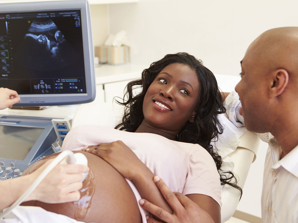
Most hospitals allow you to watch the scan, which takes about 30 minutes, as it is being carried out. If you have not had an ultrasound before now, the technician will check that there is only one baby, and will confirm your due date (Cargill and Morin 2017).
They will point out your baby's heartbeat and parts of their body, such as their face and hands, before looking at them in detail (Cargill and Morin 2017).
After you’ve seen your baby on the screen, some sonographers will turn the screen away for the rest of the scan, and show you views at the end. Some hospitals have a second monitor at the foot of the couch, so you can watch the entire scan.
Can I have a photo of my scan?
Your hospital or ultrasound clinic may be able to give you a picture of your baby. It's best to check this before you go for the appointment (Healthlink BC 2019).
It will likely be printed on thermal paper, so could fade over time. Take a picture of it on your phone so you will always have a copy.
Which differences can be seen on the scan?
Sonographers have a list of things to look out for while they are doing the scan. Most of the conditions are very rare, and some can be hard to spot at this stage. If the sonographer cannot see all they need to see, you may be called back for a follow-up scan in a few weeks’ time (PHE 2015).
- absence of the top of the head (anencephaly)
- cleft lip
- defect of the abdominal wall, where the bowel and liver protrude (exomphalos)
- defect of the abdominal wall, where the intestines protrude (gastroschisis)
- missing or very short limbs (lethal dysplasia)
- defect of the spinal cord (spina bifida)
- major kidney problems (missing or abnormal kidneys)
- hole in the muscle between the chest and the abdomen (diaphragmatic hernia)
- Trisomy 18 (Edward syndrome) or trisomy 13 (Patau syndrome), which are chromosomal abnormalities
- A bright spot on the heart, or signs of major heart problems (defects of chambers, valves or vessels) (PSO 2023)
Some conditions, such as heart defects and bowel obstructions, may not be seen until later in your pregnancy.
What if there are signs of a problem on my anatomy ultrasound?
If a scan reveals a problem, you should be referred to a specialist and get plenty of support to guide you through all of your options (GEC-KO 2018). Though serious problems are rare, in Canada, one baby in 25 is born with a condition that may need medical care or treatment (GEC-KO 2018). Some problems may mean that your baby needs treatment or surgery after birth, or while they are still in your womb.
Do I have to have an anatomy scan?
The scan provides a lot of useful information about how your pregnancy is going and if your baby is developing well, but it's your choice whether or not to have one. Early in your pregnancy, your healthcare provider should give you information about why you're being offered the scan, and what it will and will not be able to tell you. This will allow you time to decide whether you would like the scan.

Was this article helpful?
Yes
No
If the scan shows no nose bone, does my baby have Down syndrome?
By The BabyCenter Editorial TeamIf the scan shows no nose bone, does my baby have Down syndrome?
By The BabyCenter Editorial TeamIs it a problem that my baby's cord only has two blood vessels?
By Ann Elisabeth SamsonWhat is a placental lake?
By Ann Elisabeth Samson

BabyCenter's editorial team is committed to providing the most helpful and trustworthy pregnancy and parenting information in the world. When creating and updating content, we rely on credible sources: respected health organizations, professional groups of doctors and other experts, and published studies in peer-reviewed journals. We believe you should always know the source of the information you're seeing. Learn more about our editorial and medical review policies.
Audibert F, De Bie I, Johnson J et al. 2017. No 348-Joint SOGC-CCMG Guideline: Update on Prenatal Screening for Fetal Aneuploidy, Fetal Anomalies, and Adverse Pregnancy Outcomes. Journal of Obstetrics and Gynaecology Canada 1;39(9): 805 – 817.
Cargill Y, Morin L. 2017. No. 223-content of a complete routine second trimester obstetrical ultrasound examination and report. Journal of Obstetrics and Gynaecology Canada. 1;39(8):e144-9.
GEC-KO. 2018. A Guide to Understanding Prenatal Screening Tests. Genetics Education Canada -Knowledge Organization. www.geneticseducation.caOpens a new window [Accessed March 2019]
NHS. 2015. Anomaly scan at 18-21 weeks pregnant. NHS Choices, Health A-Z. www.nhs.ukOpens a new window [Accessed July 2017]
PHE. nd. Fetal anomaly screening: 18+0 to 20+6 week fetal anomaly ultrasound scan Continuing Professional Development for Screening. cpdscreening.phe.org.ukOpens a new window [Accessed July 2017]
PHE. 2015. Fetal anomaly screening programme standards: 2015 to 2016 NHS Fetal Anomaly Screening Programme. www.gov.ukOpens a new window [Accessed July 2017]
PSO. 2023. 18-22 Week Ultrasound Results Prenatal Screening Ontario [Accessed August 2023]
SOGC. nd. Routine tests. Ultrasound. Your Pregnancy. Society of Obstetricians and Gynaecologists of Canada. www.pregnancyinfo.caOpens a new window [Accessed July 2023]
TOP. 2018. Second Trimester Detailed Anatomic Study Toward Optimized Practice (TOP) Clinical Practice Guidelines for Alberta. [accessed August 2023]